Home

Cooperación trabajo neutral short axis lymph node contar Compañero explotar

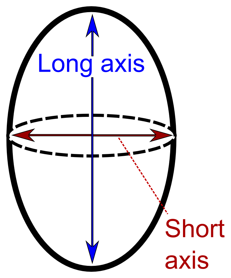
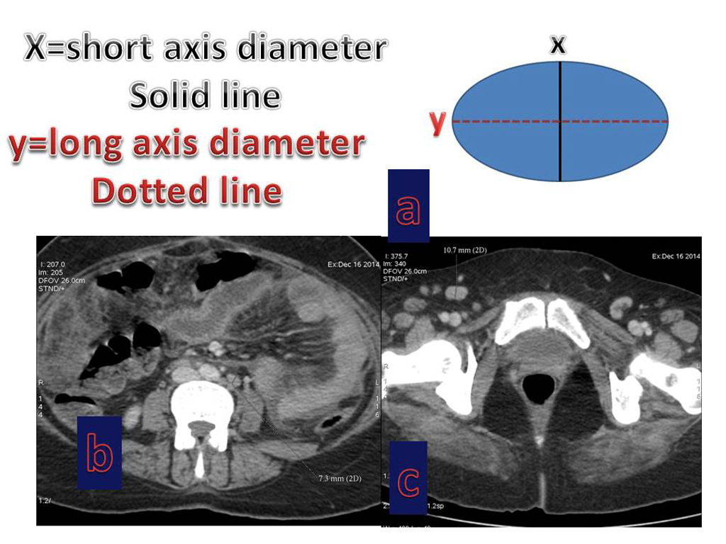
File:Long and short axis.png - Wikimedia Commons

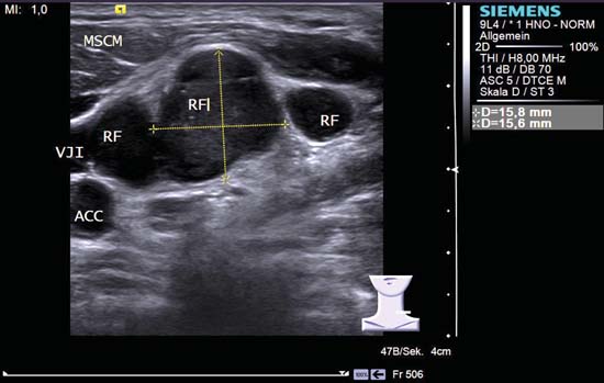
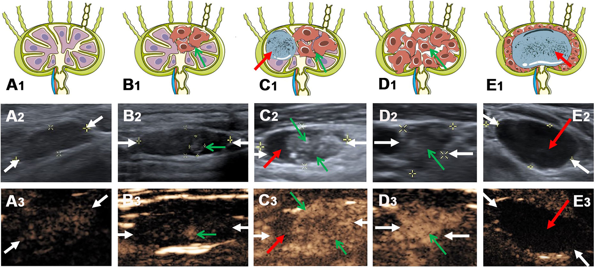
Frontiers | Value of Contrast-Enhanced Ultrasound for Evaluation of  Cervical Lymph Node Metastasis in Papillary Thyroid Carcinoma
Frontiers | Value of Contrast-Enhanced Ultrasound for Evaluation of Cervical Lymph Node Metastasis in Papillary Thyroid Carcinoma

Prediction of malignant lymph nodes in NSCLC by machine-learning  classifiers using EBUS-TBNA and PET/CT | Scientific Reports
Prediction of malignant lymph nodes in NSCLC by machine-learning classifiers using EBUS-TBNA and PET/CT | Scientific Reports

Pin on ultrasound
Pin on ultrasound

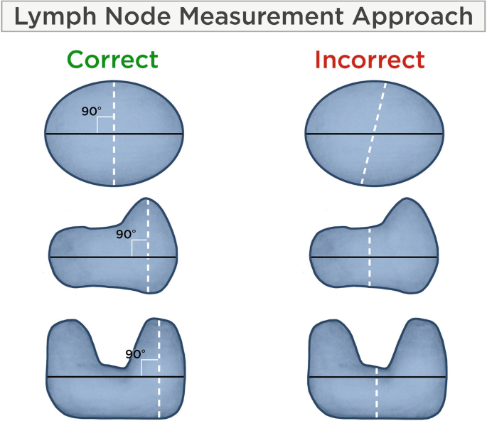
The procedure for lymph node measurement. The maximal-sized axis is... |  Download Scientific Diagram
The procedure for lymph node measurement. The maximal-sized axis is... | Download Scientific Diagram

Axillary Lymph Nodes Suspicious for Breast Cancer Metastasis: Sampling with  US-guided 14-Gauge Core-Needle Biopsy—Clinical Experience in 100 Patients |  Radiology
Axillary Lymph Nodes Suspicious for Breast Cancer Metastasis: Sampling with US-guided 14-Gauge Core-Needle Biopsy—Clinical Experience in 100 Patients | Radiology
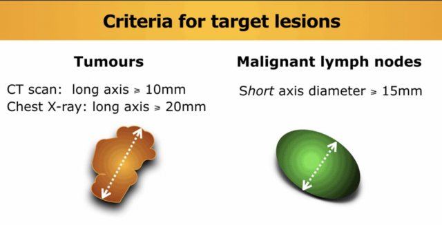
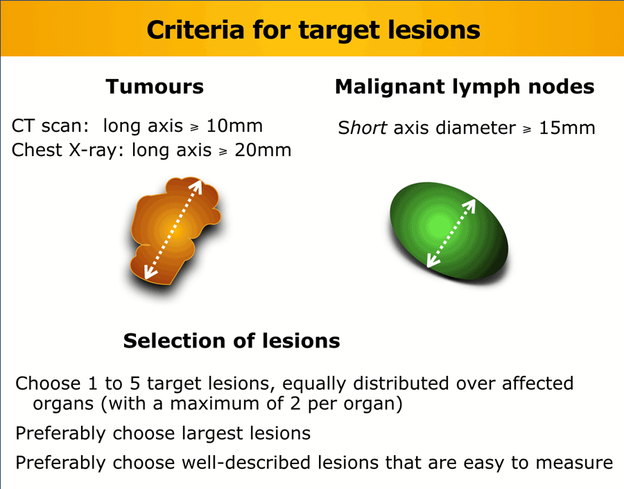
The Radiology Assistant : RECIST 1.1 - the basics

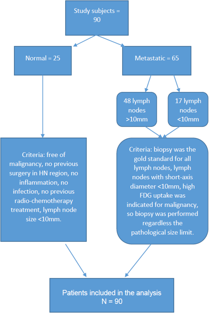
Diffusion-Weighted Imaging (DWI) derived from PET/MRI for lymph node  assessment in patients with Head and Neck Squamous Cell Carcinoma (HNSCC) |  Cancer Imaging | Full Text
Diffusion-Weighted Imaging (DWI) derived from PET/MRI for lymph node assessment in patients with Head and Neck Squamous Cell Carcinoma (HNSCC) | Cancer Imaging | Full Text
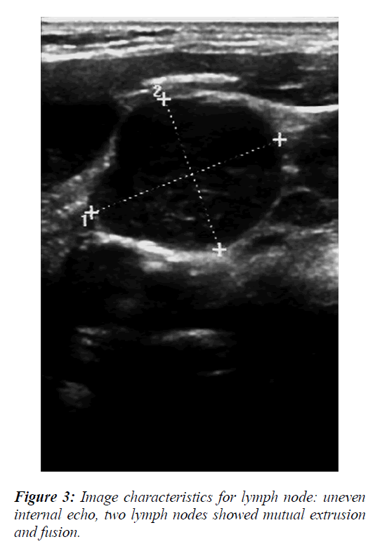
The logistic regression analysis of ultrasonographic features in  differential diagnosis of cervical lymph nodes metastasis of nasopharyngeal  carcinoma patients.
The logistic regression analysis of ultrasonographic features in differential diagnosis of cervical lymph nodes metastasis of nasopharyngeal carcinoma patients.

Lymphadenopathy - Wikiwand

Figure 9 from Lymph Node Sonography | Semantic Scholar

Drawing shows short axis of cortex (white arrow) and short axis of... |  Download Scientific Diagram
Drawing shows short axis of cortex (white arrow) and short axis of... | Download Scientific Diagram

Terms, definitions and measurements to describe sonographic features of lymph  nodes: consensus opinion from the Vulvar International Tumor Analysis  (VITA) group - Fischerova - 2021 - Ultrasound in Obstetrics & Gynecology -  Wiley Online Library
Terms, definitions and measurements to describe sonographic features of lymph nodes: consensus opinion from the Vulvar International Tumor Analysis (VITA) group - Fischerova - 2021 - Ultrasound in Obstetrics & Gynecology - Wiley Online Library
Cureus | Estimation of Accuracy of B-Mode Sonography and Elastography in  Differentiation of Benign and Malignant Lymph Nodes With Cytology as  Reference Standard: A Prospective Study | Article
Cureus | Estimation of Accuracy of B-Mode Sonography and Elastography in Differentiation of Benign and Malignant Lymph Nodes With Cytology as Reference Standard: A Prospective Study | Article

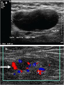
Lymphadenopathy - Wikipedia

Distribution of malignant lymph nodes based on short axis diameter (d). |  Download Table
Distribution of malignant lymph nodes based on short axis diameter (d). | Download Table

The Radiology Assistant : RECIST 1.1 - and more

A Practical Guide of the Southwest Oncology Group to Measure Malignant  Pleural Mesothelioma Tumors by RECIST and Modified RECIST Criteria -  ScienceDirect
A Practical Guide of the Southwest Oncology Group to Measure Malignant Pleural Mesothelioma Tumors by RECIST and Modified RECIST Criteria - ScienceDirect

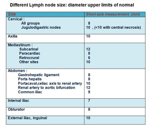
Normative Measurements of Head and Neck Lymph Nodes on Imaging |  SpringerLink
Normative Measurements of Head and Neck Lymph Nodes on Imaging | SpringerLink

EPOS™
EPOS™

B-Mode and Elastosonographic Evaluation to Determine the Reference  Elastosonography Values for Cervical Lymph Nodes
B-Mode and Elastosonographic Evaluation to Determine the Reference Elastosonography Values for Cervical Lymph Nodes
Neck Lymph Nodes | Radiology Key

Figure 2 from Lymph Node Sonography | Semantic Scholar

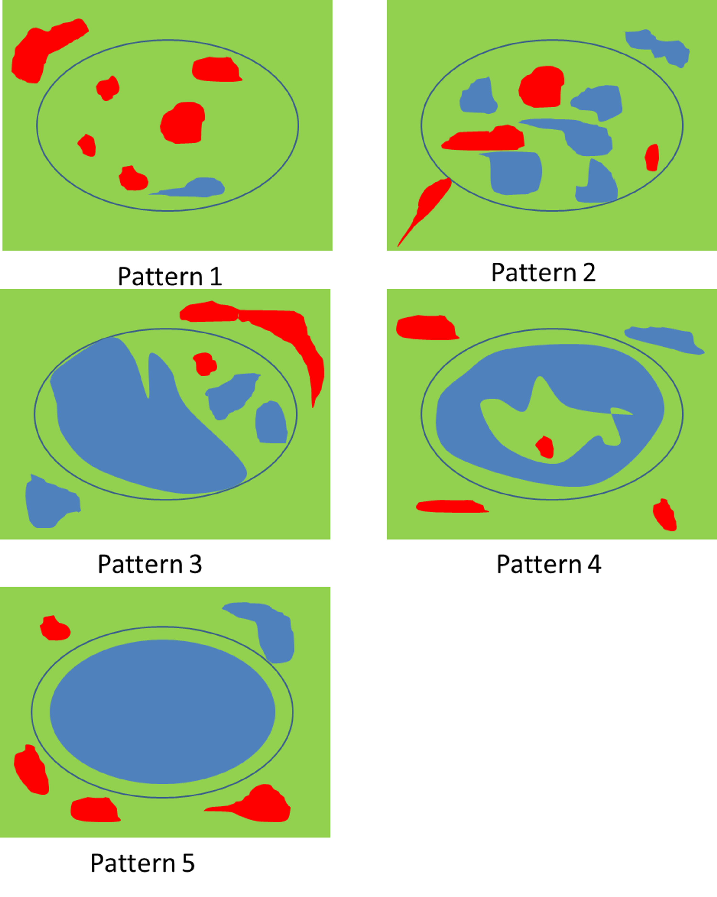
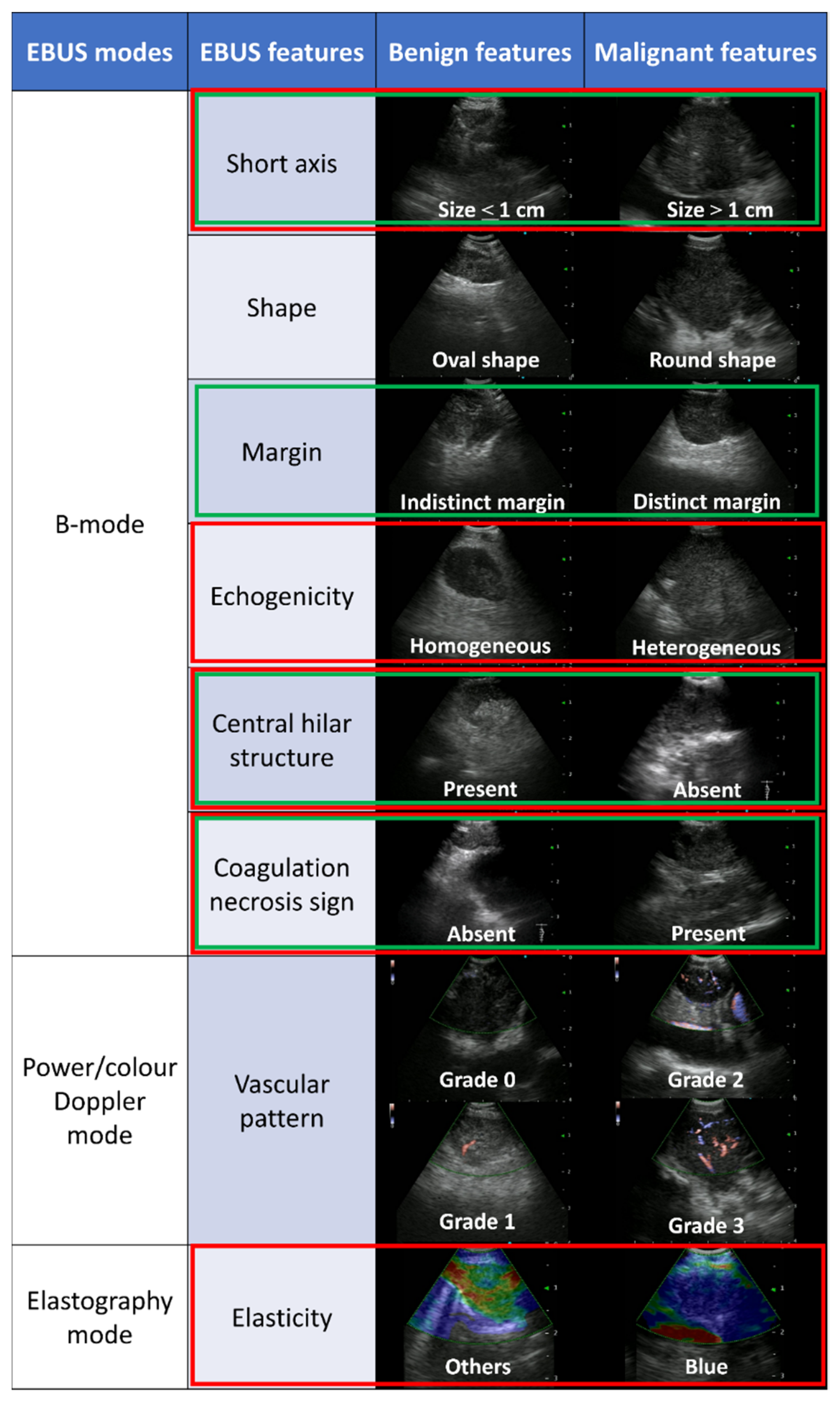
Cancers | Free Full-Text | Predicting Malignant Lymph Nodes Using a Novel  Scoring System Based on Multi-Endobronchial Ultrasound Features
Cancers | Free Full-Text | Predicting Malignant Lymph Nodes Using a Novel Scoring System Based on Multi-Endobronchial Ultrasound Features

Sonographic localisation and description of inguinofemoral lymph nodes in  patients with vulvar squamous cell carcinoma - Fenech - Sonography - Wiley  Online Library
Sonographic localisation and description of inguinofemoral lymph nodes in patients with vulvar squamous cell carcinoma - Fenech - Sonography - Wiley Online Library

EPOS™
EPOS™